Have you ever wondered what it would be like to hold a human heart in your hands? Well, thanks to the wonders of technology and 3d printing anatomical models, now you can! Prepare yourself for an exciting journey into the world of anatomy education like never before.
The Marvels of 3D Printing Anatomical Models
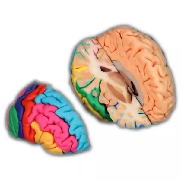
Gone are the days when students had to rely solely on textbooks and two-dimensional diagrams to understand complex anatomical structures. With the advent of 3D printing, educators have found a new tool that brings anatomy to life. These models allow students to explore intricate details and gain a deeper understanding of the human body.
Imagine being able to touch and examine a replica of a real human organ, complete with all its nooks and crannies. Students can now study these models from every angle, enhancing their spatial awareness and improving their overall comprehension.
Not only do these 3D printed anatomical models provide an immersive learning experience, but they also offer endless possibilities for customization. Educators can create replicas specific to certain medical conditions or anomalies, allowing students to practice diagnosing and treating various scenarios.
DIGIHUMAN: The Future is Here
If you thought holding a physical model was impressive enough, wait until you hear about DIGIHUMAN – an innovative project taking anatomy education by storm. This cutting-edge technology combines high-resolution imaging techniques with advanced computer algorithms to create virtual representations of real human bodies.
DIGIHUMAN allows users not only to visualize different layers within the body but also provides interactive features such as zooming in on specific areas or even virtually dissecting organs. It’s like having X-ray vision without any radioactive side effects!
With DIGIHUMAN, students can explore the human body in ways never before possible. They can navigate through intricate networks of blood vessels, examine the inner workings of organs, and even simulate surgical procedures. It’s like stepping into a virtual operating room without the pressure.
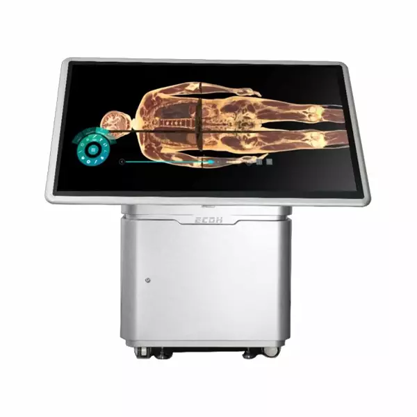
The Virtual Dissection Table: A Game-Changer
If you thought 3D printing anatomical models and DIGIHUMAN were mind-blowing, get ready for another game-changer – the virtual dissection table. This state-of-the-art technology combines augmented reality with haptic feedback to create an unparalleled learning experience.
Using this table, students can interact with digital representations of anatomical structures using touch-sensitive tools that provide realistic tactile sensations. They can feel the texture of tissues, sense resistance when cutting through layers, and even experience simulated bleeding.
The virtual dissection table not only enhances student engagement but also promotes teamwork as multiple users can collaborate simultaneously on a single model. It’s like playing a high-stakes game of Operation but with real medical knowledge at stake!
In Conclusion
Thanks to 3D printing anatomical models, projects like DIGIHUMAN, and advancements in virtual dissection tables, anatomy education has reached new heights of interactivity and immersion. Students now have access to tools that allow them to explore the intricacies of the human body in ways unimaginable just a few years ago.
Gone are the days when anatomy was merely about memorizing facts from textbooks; it has become an exciting journey filled with hands-on experiences and interactive learning opportunities. So grab your lab coat and stethoscope because it’s time to dive headfirst into this brave new world where anatomy meets technology!