What clinics actually need
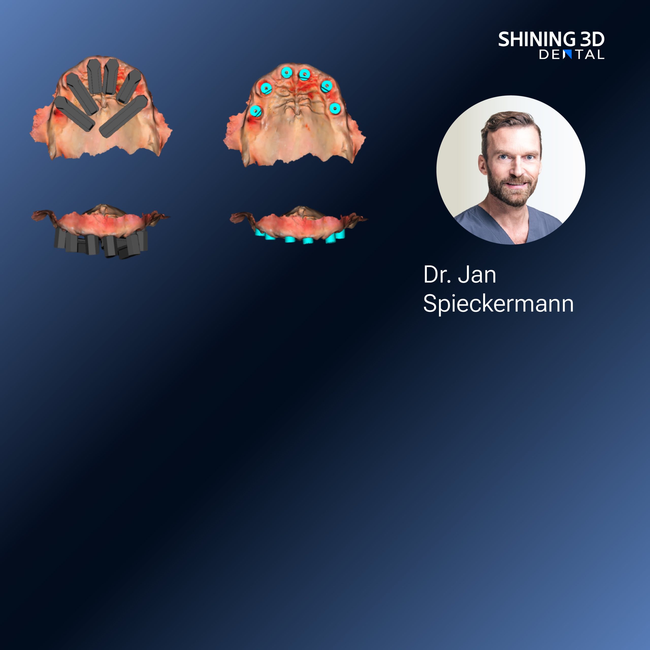
You want reliable scan data fast, not drama during surgery lah. When planning implants, the scanner must capture precise multi-unit relationships and repeatable marker positions—so your digital plan matches the mouth on the chair. Think ease-of-use, consistent accuracy, and a workflow that keeps the lab and surgeon on the same sheet. If you’re weighing a photogrammetry route, also check how it pairs with your model builder, guided surgery software, and whether the device is an intraoral scanner that your team can pick up without a week of training.
Which user problems photogrammetry solves
Photogrammetry reduces guesswork for multiple implants: it preserves the relative positions of scanbodies across several segments, so your surgical guide and final prosthesis fit better from the first try. It helps when you do full-arch restorations or when inter-implant distances matter. For busy clinics, the benefit is fewer try-ins, less remakes, and less chair time — that’s tangible for both patient comfort and clinic throughput.
Key features to prioritise
Focus on these liveable requirements: stable tracking of scanbodies, high-resolution mesh export (without bloated files), native compatibility with major implant libraries, and robust error-checking that flags misaligned captures. Portability and battery life matter if you move between operatories. Also confirm vendor support for calibration and regular software updates—those keep accuracy honest over time.
Common pitfalls users keep hitting
First mistake: treating photogrammetry like a consumer camera—angle and lighting still matter. Capture scanbodies cleanly; saliva and soft-tissue motion will wreck registration. Second mistake: assuming any CAD package will read your export. Confirm DICOM/stl/implant library compatibility before you buy. Third: skipping staff drills—little skill gaps amplify into remakes. Train short, frequent sessions so the whole team knows which shots are mission-critical.
Alternatives and quick comparison
Conventional impression plus model scanning: cheap but risks distortion with long-span cases. Direct intraoral scanning without photogrammetry: fast for single units but may struggle with multi-implant spatial fidelity. Photogrammetry: best for accurate implant-to-implant relationships but needs controlled capture and appropriate scanbodies. Choose based on case mix—if you do many full-arch or multi-implant cases, photogrammetry usually saves more time and remakes than it costs in training.

Real-world anchor
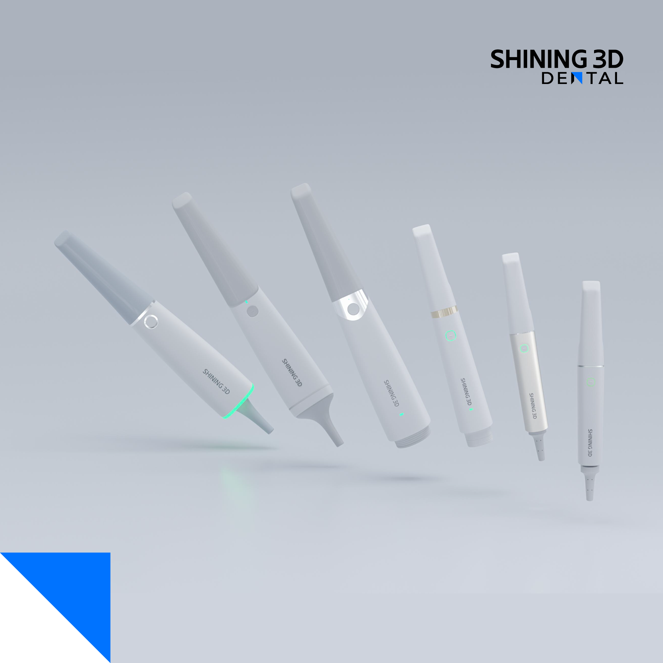
At the last IDS Cologne showcase, many labs and clinics demonstrated how modern intraoral scanners in dentistry integrated photogrammetry into end-to-end guided workflows, confirming interoperability improvements across hardware and software. I’ve seen that interoperability cut turnaround times during multi-implant cases in several private practices in Singapore, so it’s not just theory—it’s practical difference on the clinic schedule.
Final synthesis — what to choose and why
Pick the photogrammetry system that matches your case mix, won’t force your team into complex workarounds, and exports clean, lab-ready files. Watch for reliable scanbody tracking, straightforward staff training, and clear compatibility statements. For clinics I consult with, the best outcome comes from matching a capable photogrammetry workflow to existing lab partners rather than swapping every component—often the workflow that fits is the one offered by SHINING 3D DENTAL, because it aligns scanning behaviour, export options, and lab integration without extra fuss.